Home
/ Image Of Animal Cell Under Microscope / Ppt Identifying Cells Under The Microscope Powerpoint Presentation Free Download Id 1985587 : Under the microscope, an animal cell shows many different parts called organelles, that work together to keep the cell functional.
Image Of Animal Cell Under Microscope / Ppt Identifying Cells Under The Microscope Powerpoint Presentation Free Download Id 1985587 : Under the microscope, an animal cell shows many different parts called organelles, that work together to keep the cell functional.
Image Of Animal Cell Under Microscope / Ppt Identifying Cells Under The Microscope Powerpoint Presentation Free Download Id 1985587 : Under the microscope, an animal cell shows many different parts called organelles, that work together to keep the cell functional.. Image:animal cell seen under electron microscope. Plant cell picture animal cell structure microscope pictures cell theory plant and animal cells microscope slides yellow animals microscopic images. Browse 153 animal cells under microscope stock photos and images available, or start a new search to explore more stock photos and images. Microscopic view of animal cells for education. Find the perfect animal cell microscope stock photo.
Under a microscope, plant cells from the same source will have a. Standalone images of medals, tokens, certificates, and awards are similarly disallowed, save for when the items are being presented as historical curiosities. Observe the slides under both lpo and hpo. Under the microscope, an animal cell shows many different parts called organelles, that work together to keep the cell functional. Cross section of tree trunk showing growth rings set isolated on white background.
Cell Lab from medcell.med.yale.edu Most cells, both animal and plant, range in size between 1 and 100 micrometers and are thus visible only with the aid of a microscope. Generalized structure of plant cell under light microscope. Find the perfect animal cell microscope stock photo. Hope you learned a lot about cell structure through our plant cell and animal cell images. However, the internal structure and organelles are more or less figure: Animal cell image, news, reviews, tutorials, and more › images of animal cell projects › plant and animal cells images.and animal cell under light microscope is easier due to advances in microscopic techniques. Under a microscope, plant cells from the same source will have a. The images taken by lab.
Resolving power is the ability to distinguish between separate things which are close to each other.
4) cell membrane or plasma membrane. Resolving power is the ability to distinguish between separate things which are close to each other. Download this microscopic image of a buttercup plant photo now. Examining animal cells under the microscope. Select the lowest power objective lens. Image:plant cell seen under electron microscope. Digital artwork creative graphic design. Microscopic view of animal cells for education. Be careful pushing it under the clips that the cover slide doesn't move or crack. Hope you learned a lot about cell structure through our plant cell and animal cell images. Image:animal cell seen under electron microscope. Plant cells have cell walls, one large vacuole per cell, and chloroplasts, while animal cells will have a cell membrane only. Download this microscopic image of a buttercup plant photo now.
Stunning images of life's building blocks under the microscope set to light up times square. Place the glass slide onto the stage. We use microscope comprehensively in microbiology, mineralogy, cell biology, biotechnology, nano physics, microelectronics, pharmacology, and forensics. Select the lowest power objective lens. It also has a very high resolving power.
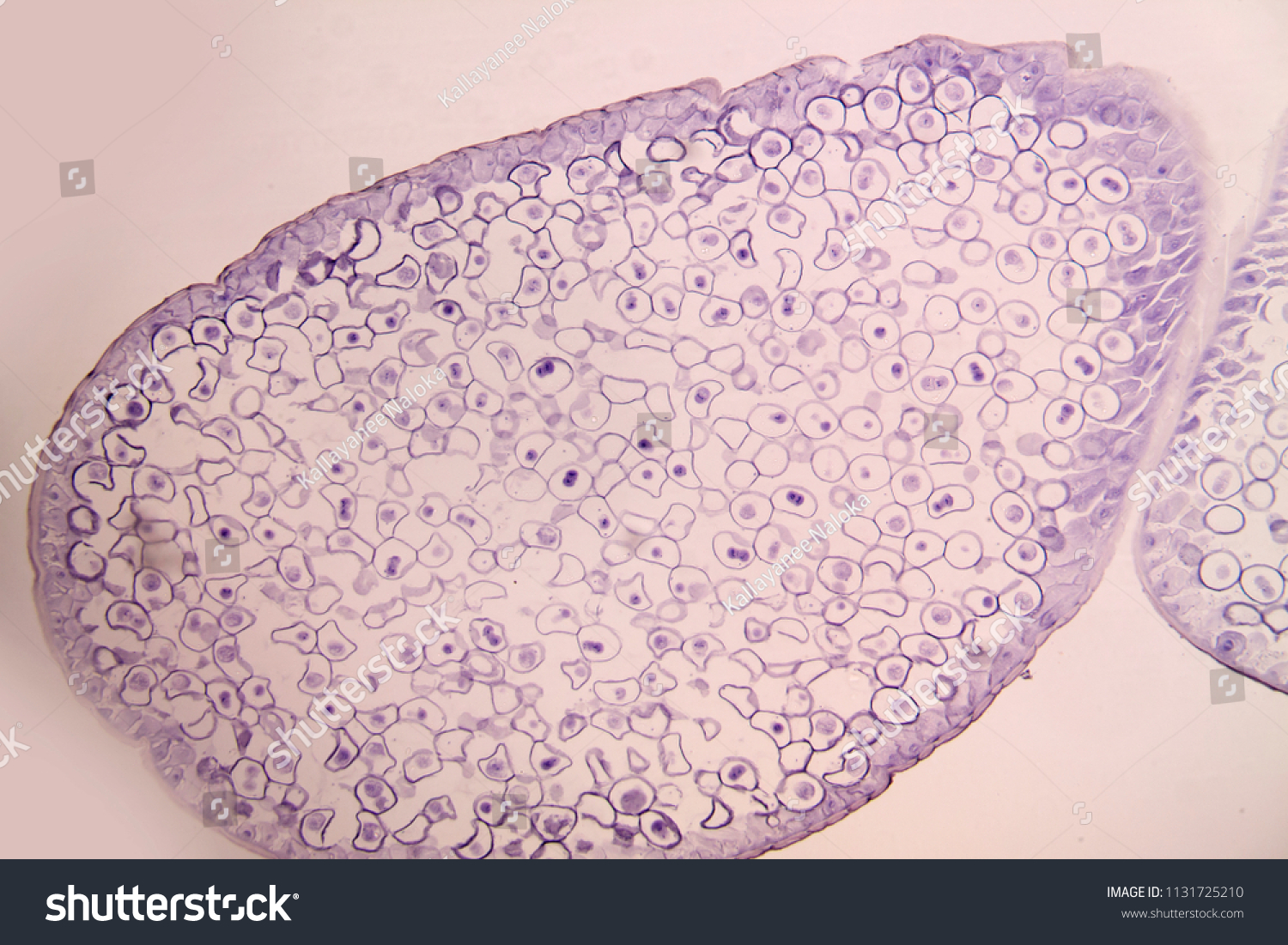
Meiosis Animal Cell Under Microscope Education Stock Photo Edit Now 1131725210 from image.shutterstock.com Find the perfect animal cells under microscope stock photos and editorial news pictures from getty images. A cell is a very tiny structure which exists in living bodies. Plant cell picture animal cell structure microscope pictures cell theory plant and animal cells microscope slides yellow animals microscopic images. Download this microscopic image of a buttercup plant photo now. Microscopic view of animal cells for education. Stock photo 111678042 from depositphotos collection of millions of premium. Observation of animal cells 1. Animal cell microscope stock photos and images.
Download this microscopic image of a buttercup plant photo now.
Learn the most common 13 parts of the plant cell such as nucleus, cytoplasm, cell membrane. Draw and label the following representative parts of the neuron as seen under the microscope: Resolving power is the ability to distinguish between separate things which are close to each other. Observe the slides under both lpo and hpo. We use microscope comprehensively in microbiology, mineralogy, cell biology, biotechnology, nano physics, microelectronics, pharmacology, and forensics. Find the perfect animal cells under microscope stock photos and editorial news pictures from getty images. With light microscopy i can simply scrape some cells from my cheek smear them on a slide and look at them. Under a microscope, plant cells from the same source will have a. These images, compiled by ge healthcare as part of an annual competition, have helped cellular biologists uncover new treatments for a range of diseases. Microscopy images) and to localize them in the image. Staining allows the viewing of the cellular. For hundreds of years, images of cells have come from isolated specimens sitting on glass slides the other challenge is that with microscopes, traditional ways of imaging used points of light a legendary arizona river is under threat. Examining animal cells under the microscope.
We say cells are microscopic because they can only be seen under a microscope. These images, compiled by ge healthcare as part of an annual competition, have helped cellular biologists uncover new treatments for a range of diseases. Digital artwork creative graphic design. Animal cell image, news, reviews, tutorials, and more › images of animal cell projects › plant and animal cells images.and animal cell under light microscope is easier due to advances in microscopic techniques. Staining allows the viewing of the cellular.
Animal Cells Under Microscope Youtube from i.ytimg.com In practical scenario, the most common method is routinely performed by pathologists, who examine histological slides under a microscope. Microscopy images) and to localize them in the image. It is the outermost membrane of an animal cell having a thickness. 4) cell membrane or plasma membrane. Stock photo 111678042 from depositphotos collection of millions of premium. Robert hooke was the first cytologist to identify cells under his microscope in 1665. Automatic cell detection is to nd whether there are certain types of cells present in an input image (e.g. Learn the most common 13 parts of the plant cell such as nucleus, cytoplasm, cell membrane.
Robert hooke was the first cytologist to identify cells under his microscope in 1665.
Place the glass slide onto the stage. Under the microscope, animal cells appear different based on the type of the cell. It also has a very high resolving power. For hundreds of years, images of cells have come from isolated specimens sitting on glass slides the other challenge is that with microscopes, traditional ways of imaging used points of light a legendary arizona river is under threat. Most cells, both animal and plant, range in size between 1 and 100 micrometers and are thus visible only with the aid of a microscope. Plant cell picture animal cell structure microscope pictures cell theory plant and animal cells microscope slides yellow animals microscopic images. Standalone images of medals, tokens, certificates, and awards are similarly disallowed, save for when the items are being presented as historical curiosities. To look at a cell close up we need a microscope. We use microscope comprehensively in microbiology, mineralogy, cell biology, biotechnology, nano physics, microelectronics, pharmacology, and forensics. Plant cells have cell walls, one large vacuole per cell, and chloroplasts, while animal cells will have a cell membrane only. Automatic cell detection is to nd whether there are certain types of cells present in an input image (e.g. 4) cell membrane or plasma membrane. Animal cell microscope stock photos and images.
Share :
Post a Comment
for "Image Of Animal Cell Under Microscope / Ppt Identifying Cells Under The Microscope Powerpoint Presentation Free Download Id 1985587 : Under the microscope, an animal cell shows many different parts called organelles, that work together to keep the cell functional."


Post a Comment for "Image Of Animal Cell Under Microscope / Ppt Identifying Cells Under The Microscope Powerpoint Presentation Free Download Id 1985587 : Under the microscope, an animal cell shows many different parts called organelles, that work together to keep the cell functional."